The Nervous System: Structure, Function, and Development
1. Evolution of the Nervous System
The first single-celled animals, or protozoa, were primitive and exhibited irritability. When evolution led to the emergence of multicellular organisms, or metazoans, specialized cells appeared for intercellular communication. These cells were responsible for the organisms’ irritability. The main function of this early nervous system was the perception of stimuli and the generation of simple responses. As the nervous system evolved, a more sophisticated communication network, the “peripheral nervous system,” extended throughout the body, along with a coordination center, the “central nervous system.” Worms developed a cord-like nervous system running along their body with a large nerve center, or ganglion, containing neurons in the anterior region. This was likely the forerunner of the vertebrate brain and spinal cord. A significant development in evolution was the appearance of afferent and efferent nerves in the peripheral nervous system. As animals evolved, they acquired eyes, olfactory receptors for smell, and hearing organs in the anterior part of their bodies. The brain appeared early on as a small swelling, and it underwent significant development as complexity increased. Evolution added new structures onto the existing ones, such as the brainstem, spinal cord, and cerebellum. The nervous system’s evolution brought about flexibility and adaptability in responding to stimuli as environmental pressures grew.
Invertebrates are animals without a nervous system capable of complex, real-mind behaviors.
The vertebrate spinal cord, while not as developed as the brain, is the center of reflex behavior in all vertebrates. It conducts impulses between sensory organs and muscles on one side of the body and the brain on the other. However, this doesn’t mean that the spinal cord hasn’t undergone changes throughout evolution. Spinal nerves regulate lower bodily functions in both fish and humans. In fish, there are autonomous complexes like sympathetic ganglia with a distinct autonomic system.
2. Embryology
Segmentation: 2-16 cells
16 to 60 cell morula
Divide into two groups of cells:
- Trophoblasts that give rise to the placenta, responsible for nutrition
Blastula
Cells divide into three groups:
- Ectoderm: Nervous system
- Mesoderm: Urinary system
- Endoderm: Gut, liver, pancreas
Invaginates into neural tube and neural crest
Neural tube gives rise to the central nervous system, comprising the brain and spinal cord
Neural crest gives rise to:
- Afferent or sensory neurons (dorsal region)
- Efferent or motor neurons (ventral region)
Neural tube forms three primary vesicles and then five secondary vesicles:
- Prosencephalon:
- Telencephalon: Cerebral hemispheres
- Diencephalon: Thalamus and hypothalamus
- Mesencephalon: Quadrigeminal plate, cerebral peduncles
- Rhombencephalon: Pons, cerebellum, medulla oblongata
- Myelencephalon: Spinal cord
- Prosencephalon:
3. Neurons
Neurons are specialized cells in the nervous system characterized by the excitability of their plasma membrane. They are responsible for receiving stimuli and conducting nerve impulses (in the form of action potentials) to other neurons or target cells like muscle fibers. Highly differentiated, most neurons do not divide after reaching maturity, although a small percentage do. Neurons have typical morphological characteristics that support their functions: a central cell body or ‘perikaryon’; one or more short extensions called dendrites, which usually transmit impulses toward the cell body; and a long extension called an axon or ‘axis cylinder’, which conducts impulses away from the cell body to another neuron or target organ.
Neurogenesis, the creation of new neurons, in adult beings was only discovered in the latter part of the 20th century. Until recent decades, it was believed that, unlike most other body cells, mature neurons in the individual did not regenerate, except for olfactory cells. Myelinated nerves of the peripheral nervous system also have the ability to regenerate through the use of the neurolemma, a layer formed from the nuclei of Schwann cells.
Nucleus
Located in the cell body, the nucleus usually occupies a central position and is very conspicuous (visible), especially in small neurons. It contains one or two prominent nucleoli and dispersed chromatin, suggesting the relatively high transcriptional activity of this cell type. The nuclear envelope, with plenty of nuclear pores, has a well-developed nuclear lamina. Within the nucleus, you may find the Cajal body, a spherical structure about 1 micron in diameter that corresponds to an accumulation of protein rich in the amino acids arginine and tyrosine.
Perikaryon
Several organelles fill the cytoplasm surrounding the nucleus. The most notable organelle in the perikaryon is the Nissl substance, which is filled with ribosomes attached to free and rough endoplasmic reticulum. Under a light microscope, it appears as basophilic clumps, and under an electron microscope, as stacks of endoplasmic reticulum cisternae. Such an abundance of organelles related to protein synthesis is due to the high biosynthetic rate of the perikaryon.
These are particularly notable in somatic motor neurons, as in the anterior horn of the spinal cord or certain cranial nerve motor nuclei. Nissl bodies are found not only in the perikaryon but also in dendrites, but not in the axon, which is one way to differentiate dendrites and axons in the neuropil.
The Golgi apparatus, which was originally discovered in neurons, is a highly developed system of flattened and small agranular vesicles. It is the region where the products of the Nissl substance undergo further synthesis. Primary and secondary lysosomes (the latter rich in lipofuscin, which may marginalize the nucleus in older individuals due to its large increase) are also present. The mitochondria, small and rounded, usually possess longitudinal ridges.
As for the cytoskeleton, the perikaryon is rich in microtubules (classically called neurotubules, although they are identical to those of non-neuronal cells) and intermediate filaments (called neurofilaments). The neurotubules are involved in the rapid transport of protein molecules that are synthesized in the cell body and are carried through the dendrites and axon.
Dendrites
Dendrites are cytoplasmic projections branching from the neuronal soma, enveloped by a membrane without myelin. They sometimes have an irregular contour, developing spines. Their characteristic organelles and components include: many microtubules and few neurofilaments, both arranged in parallel bundles; many mitochondria; clumps of Nissl substance, most abundant in the area adjacent to the soma; and smooth endoplasmic reticulum, especially in the form of vesicles associated with synapses.
Axon
The axon is an extension of the neuronal soma coated by one or more Schwann cells in the peripheral nervous system of vertebrates, producing myelin or not. It can be divided, moving centrifugally from the perikaryon, into: the axon hillock, the initial segment, and the rest of the axon.
Axon hillock: Adjacent to the perikaryon, it is very visible in large neurons. It shows the gradual disappearance of the Nissl clumps and an abundance of microtubules and neurofilaments that are organized into parallel bundles in this region and will be extended along the axon.
Initial segment: This is where external myelination begins, if present. In the cytoplasm at this point, there is an electron-dense area rich in material continuous with the plasma membrane, composed of filamentous material and dense particles. It is assumed to play a role in the generation of action potentials that transmit the synaptic signal. As for the cytoskeleton, this area has the same organization as the rest of the axon. The microtubules are polarized and have the τ protein but not the MAP-2 protein.
Rest of the axon: In this section, we begin to see the nodes of Ranvier and synapses.
Classification
Although the cell body size can range from 5 to 135 micrometers, extensions or dendrites can span a distance of over a meter. The number, length, and branching pattern of dendrites provide a method for the morphological classification of neurons.
According to the size of the extensions, neurons are classified as:
Polyhedral: Like the motor neurons of the anterior horn of the spinal cord.
Spindle: Like double bouquet cells in the cerebral cortex.
Star: Like arachniform and stellate neurons of the cerebral cortex and stellate, basket, and Golgi cells in the cerebellum.
Spherical: In dorsal root ganglia, sympathetic and parasympathetic ganglia.
Pyramidal: Present in the cerebral cortex.
According to polarity:
Monopolar or unipolar neurons: These have only one extension that forks and behaves functionally as an axon. However, the branches at their ends in the periphery receive signals and function as dendrites, transmitting the impulse without it passing through the neuronal soma. They are typical of invertebrate ganglia and the retina.
Bipolar neurons: They possess a cell body, an elongated dendrite, and an axon (there can be only one axon per neuron). The nucleus of this type of neuron is located in the center, so it can send signals to both poles. Examples of these neurons are found in the bipolar cells of the retina (cones and rods) and the cochlear and vestibular ganglia, which are specialized for receiving sound and balance information.
Multipolar neurons: They have a large number of dendrites arising from the cell body. These cells are the classic neuron with small extensions (dendrites) and a long extension, or axon. They represent the majority of neurons. Within the multipolar category, we distinguish between Golgi type I neurons, which have a long axon, and Golgi type II neurons, which have no axon or a very short one. Projection neurons are of the first type, and local neurons or interneurons are of the second type.
Pseudounipolar neurons: These have a single dendrite or neurite that divides at a short distance from the cell body into two branches, hence the name pseudounipolar (pseudo meaning”fals”). One branch is directed toward a peripheral structure, and the other enters the central nervous system. Examples of this type of neuron are found in the dorsal root ganglia.
Anaxonic neurons: These are small neurons where dendrites are indistinguishable from axons. They are found in the brain and special sense organs.
According to the characteristics of neurites:
According to the nature of the axon and dendrites, neurons are classified into:
Golgi type I neurons: These have a very long axon that branches away from the perikaryon. Axons can be up to 1 meter long.
Golgi type II neurons: These have a short axon that branches near the cell body.
Neurons without a defined axon: Such as amacrine cells of the retina.
Isodendritic neurons: These have straight dendrites that branch so that the daughter branches are longer than their parent branches.
Idiodendritic neurons: These have dendrites organized depending on the neuronal type, such as the Purkinje cells of the cerebellum.
Allodendritic neurons: These are intermediate between the two previous types.
According to the chemical mediator:
Neurons can be classified according to the chemical mediator they release:
Cholinergic: Release acetylcholine.
Noradrenergic: Release norepinephrine.
Dopaminergic: Release dopamine.
Serotonergic: Release serotonin.
GABAergic: Release GABA, or γ-aminobutyric acid.
4. Synapses
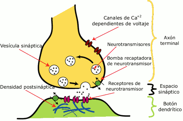
The synapse (from Gr. σύναψις,”lin”) is the process of communication between neurons. It begins with a chemical signal that causes an electrical discharge in the presynaptic cell membrane (the sending cell). Once the nerve impulse reaches the end of the axon, the neuron secretes a type of protein (neurotransmitters) that are deposited in the synaptic cleft, the space between the presynaptic neuron and the postsynaptic neuron (the receiving cell). These neurotransmitters are responsible for exciting or inhibiting the action of another neuron. 
5. Neurotransmitters
A neurotransmitter is a biomolecule, usually synthesized by neurons, that is released from vesicles in the presynaptic neuron into the synaptic cleft, causing a change in the action potential of the postsynaptic neuron. Neurotransmitters are, therefore, the primary signaling molecules in the synapse.
6. Action Potentials
An action potential, also called an electrical impulse or spike, is a wave of electrical discharge that travels along the cell membrane. Action potentials are used in the body to carry information between tissues, making them a crucial microscopic process essential to the life of animals. They can be generated by different types of body cells, but the most active in their use are the cells of the nervous system, which use them to send messages between nerve cells or from nerve cells to other body tissues such as muscle or glands.
Many plants also generate action potentials that travel through the phloem to coordinate their activity. The main difference between action potentials in animals and plants is that plants use flows of potassium and calcium, while animals use potassium and sodium.
Action potentials are the primary means of transmission of neural codes. Their properties allow for rapid signaling over long distances within the body, enabling centralized control and coordination of organs and tissues.
Threshold and Initiation
Action potentials are triggered when an initial depolarization reaches a threshold. This threshold potential varies but is usually around -55 to -30 mV relative to the resting potential of the cell, which implies that the influx of sodium ions exceeds the outflow of potassium ions. The net flow of positively charged sodium ions accompanying depolarization further depolarizes the membrane potential, resulting in the opening of voltage-gated sodium channels. These channels provide a greater flow of inward ionic currents, further increasing the depolarization in a positive feedback loop that causes the membrane depolarization to reach high levels.
The threshold of the action potential can be varied by changing the balance between sodium and potassium currents. For example, if some of the sodium channels are inactive, a greater level of depolarization is required to open more sodium channels, and thus the depolarization threshold required to initiate the action potential increases. This is the principle of operation of the refractory period.
Action potentials are highly dependent on the balance between sodium and potassium ions (although other ions contribute a minority of the potential, such as calcium and chlorine), and therefore models are made with only two transmembrane ion channels: a voltage-gated sodium channel and a potassium leak channel. The origin of the action potential threshold can be viewed on the I/V curve, which represents the ionic currents through channels versus membrane potential. (The I/V curve displayed in images is an instantaneous relationship between currents. It shows the peak currents given a voltage, recorded before any inactivation occurs (1 ms after reaching this voltage for sodium). It is also important to note that the most positive voltages on the chart can only be achieved by artificial means, by applying electrodes to the membranes).
The action potential threshold is sometimes confused with the threshold of the opening of sodium channels. This is a misnomer since sodium channels do not have a threshold. Instead, they open in response to depolarization randomly. Depolarization does not mean both the opening of the channels and the increased probability of opening. Even at hyperpolarized potentials, a sodium channel can open sporadically. Additionally, the action potential threshold is the voltage at which the flow of sodium ions becomes significant, that is, the point where it exceeds the flow of potassium.
Biologically, in neurons, depolarization originates from dendritic synapses. In principle, action potentials could be generated at any point along the nerve fiber. When Luigi Galvani discovered animal electricity by making the leg of a dead frog twitch by touching the sciatic nerve with a scalpel, he was inadvertently applying an electrostatic charge and initiating an action potential.
7. Microglia
Microglia are cells with small, elongated nuclei and short, irregular extensions that have phagocytic capacity. Their precursors originate in the bone marrow and reach the nervous system through the blood, making up the mononuclear phagocyte system in the central nervous system.
They contain lysosomes and residual bodies. Generally, microglia are classified as neuroglia cells. They express the common leukocyte antigen and class II histocompatibility antigen, characteristic of antigen-presenting cells.
